|
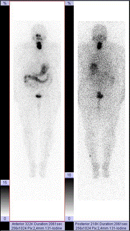
| Figure 3:Radioiodine imaging of thyroid cancer metastases using rTSH stimulation, i.m. (Thyrogen) 1 and 2 days before WB imaging at 45 h p.i.. The administered oral acivity was 1.056 GBq. This patient had papillary carcinoma of thyroid gland pT2N0M0, no invasion to capsule or lymph nodes. Whole body imaging demonstrates a quite large thyroid residue in both lobes and in the midline (12% of WB activity at 2 d), but no suspicious foci outside the neck region in planar imaging. Normal physiologic activity is seen in nasopharynx, salivary glands, stomach, GI tract and urinary bladder. A minor skin contamination is seen laterally in the left calf. |