|
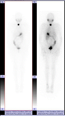
| Figure 4:Radioiodine imaging of thyroid cancer metastases using thyroxin withdrawal for 4 weeks. WB imaging was performed at 70 h p.i.. The administered oral acivity was 3.632 GBq. This patient had follicular carcinoma of thyroid gland pT1NxMx, no invasion to capsule or lymph nodes. Whole body imaging demonstrates a small thyroid residue in the right lobe (3% of WB activity at 3 d), and no suspicious foci outside the neck region in planar imaging. Normal physiologic activity is seen in salivary glands, stomach, GI tract and urinary bladder. A minor skin contamination is seen in the left ankle. |