|
||
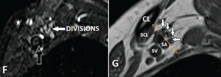
| Figure 2F and 2G: Normal MR imaging planes and appearance of BP. F-G, At the level of crossing of clavicle (star, CL) in the costoclavicular space (yellow line in between the CL and first rib), oblique sagittal fat-suppressed T2-weighted (F) and T1-weighted (G) image show the multiple nerve plexus as divisions (white arrows), superior to subclavian vessels (open arrow, SA-subclavian artery and SV-subclavian vein), CL- clavicle, SCL- subclavius muscle | ||