 |
||
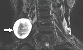
| Figure 4: A 75-year-old male with schwannoma. Coronal fat-suppressed T2-weighted MR image shows a rounded mass with central low signal and peripheral high signal intensity (target sign) (solid arrow) along the C5 postganglionic spinal nerve root with a well defined hypointense capsule. Note the nerve (open arrow) is eccentric in relation to the mass. | ||