 |
||
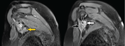
| Figure 9: A 54-year-old female on follow up for carcinoma right breast with focal infiltration of BP by contiguous spread from metastatic axillary adenopathy. Oblique sagittal fat-suppressed post contrast T1-weighted contiguous MR images show multiple enhancing axillary nodal metastasis (yellow arrow) with extracapsular spread and infiltration of pectoralis minor muscle (black arrow). There is also encasement of axillary vessels (open arrow) and infiltration of the cords of the BP (white arrow). | ||