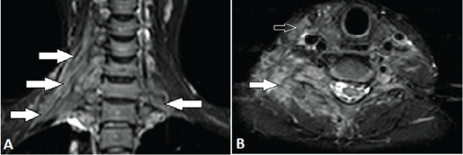
hyperintensity and enhancement (not shown) along the BP (arrows). Note the clumping of plexus elements on the right side with loss of internal architecture. B, Axial fat-suppressed T2-weighted MR image shows high signal intensity in the sternocleidomastoid (open arrow), scalene and paraspinous muscles (white arrow) on the right side and adjacent soft tissue of the neck.