 |
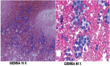
| Figure 3: Giemsa 10 X and 40 X zoom images of the slides of the FNAC sample from the nodule in the right lobe, showing moderately cellular showing sheets, papillaroid clusters, repetative follicles and singly dispersed epithelial cells exhibiting mild anisocytosis and finely granular chromatin and mild degree of nuclear overlapping. Occasional cells have longitudinal grooves. No definite intranuclear inclusions. Hurthle cells are also noted in clusters and singly dispersed. |