|
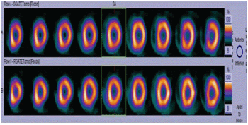
| Figure 4: Stress and Rest 99mTc- MIBI Myocardial Perfusion scintigraphy axial images (99mTc- MIBI, high resolution collimators, 64 x 64 matrix, 1.45 x zoom, both detectors, 37 views, detector configuration 76 degree, non-circular orbit, step and shoot mode acquisition) rom apex to base of the heart showing nor-mal perfusion in all segments of the left ventricular myocardium with no evidence of ischemia. Stress gated images were acquired after 45 min of 99mTc- MIBI injection at peak exercise (after the patient had achieved >85% target heart rate and double product of >25,000).Rest gated images were acquired after 45 min of reinjection of 30 mCi 99m Tc- MIBI at rest. |