|
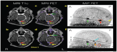
| Figure 1: Post-contrast T1-weighted MRI images alone (1a-b, left) and fused with 68Ga-DOTATOC PET (1c-d, middle) and maximum image projections (MIP) (1e-f, right) at stereotactic radiotherapy planning (1a, 1c, 1e, top) and after 11 months (1b, 1d, 1f, bottom). The MIP shows all activity in the head in the sagittal plane. The broken lines in the MIP show the relative locations of slices 1-3 and colored arrows the lesions in Figures 1 and 2. The MIP (1e) shows the position of slice 1 with only 2 active foci, the recurrent tumor (red arrow and circle) and physiological 68Ga- DOTATOC uptake in the pituitary gland (green arrow), respectively. Initial MRI (1a) indicated a 5 mm in diameter regional tumor recurrence (red arrow) that was confirmed with 68Ga- DOTATOC PET (1c). 68Ga-DOTATOC PET was used for radiotherapy planning and MRI and PET 11 month later (1b and 1d) showed structural and functional response on MIP (1f, empty red circle). Initially a larger en plaque meningioma was suspected on MRI (1a, yellow arrow) but this could not be confirmed on PET (1c) indicating reactive changes and the area was not included in the therapy field. This area was structurally and functionally stationary on follow-up. Orientation: R: Right; L: Left; A: Anterior; P: posterior. |