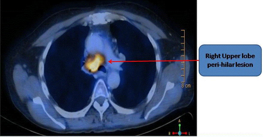
Figure 2:
Baseline PETCT scan showing FDG avid right upper lobe perihilar lesions, mediastinal lymphdenopathy.