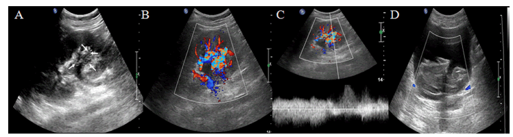
1A: A longitudinal scan with the patient in left anterior oblique position demonstrate a slightly hyperechoic structure located the inferiorcalyx
of the left kidney, with unclear boundary and tubular anechoic internal echo (white arrows).
2B: Color Doppler ultrasound imaging revealed high velocity with aliasing and continuous turbulent flows were in the lesion.
3C: Pulsed Doppler ultrasound imaging showed turbulent high-velocity flow spectrum with a burr-like boundary.
4D: Bladder ultrasound imaging showed a lot of blood clot located in the bladder cavity.