|
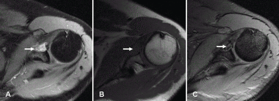
| Figure 2: MRI of left shoulder joint showing axial soft tissue lesion measuring ~18 ×15 ×15 mm anterior to the glenoid of scapula on left side. The lesion appears hypointense on T1 images (A and B) and shows mixed signal on T2 images (C) with multiple dark foci with homogenous enhancement on post contrast study (A), suggestive of pigmented villonodular synovitis arising from the subcapsular bursa. |