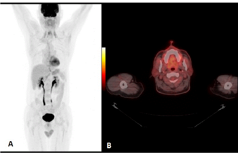
Figure 2:
A. Maximum intensity projection (MIP) image and B. axial slice of repeated FDG PET/CT scan. Normal physiologic FDG distribution is seen.