|
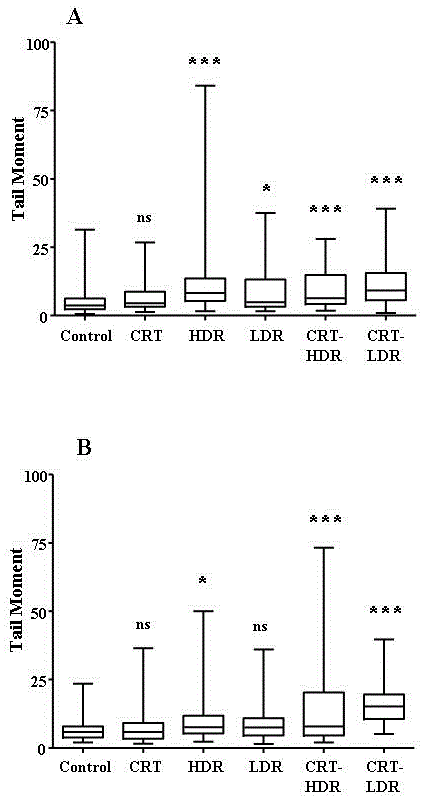
| Figure 5: Assessment of DNA damage at (A) 1 h and (B) 24 h by single cell gel electrophoresis. 100 cells were assayed per treatment. For each treatment, boxplots represent the 25th percentile, median and 75th percentile. Whisker bars show the minimum and maximum values observed. Statistical significance: one (*) and three (***) symbols denote p<0.05 and p<0.0001 respectively. |