 |
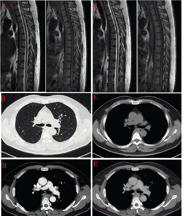
| Figure 1: The first sagittal spinal column MRI scan of T2WI and T1WI image (A). In the chest CECT scan, the lung and mediastinal windows of plain scan images (B and C). In the chest CECT scan, the enhanced images (D and E). The second follow-up sagittal spinal column MRI scan (F). |