 |
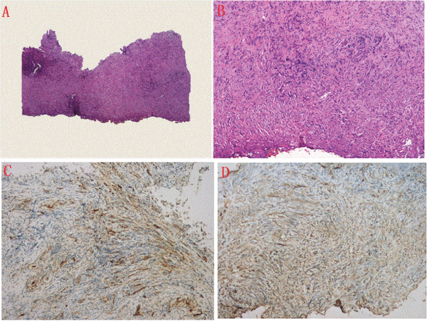
| Figure 3: The photomicrographs of the biopsies for the mass (A, hematoxylin-eosin, original magnification 40×; B, hematoxylineosin, original magnification 100×). The immunohistochemistry results that the mass had positive for CD34 and CD99 (C and D). |