|
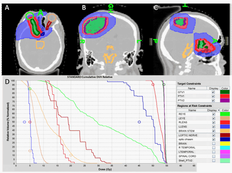
| Figure 2: Tomoplan of a patient with two separate lesions treated simultaneously in one single plan. Dose wash of 54Gy (green), 50Gy (red), and 30Gy (blue) is displayed showing excellent target volume coverage of right optic nerve sheath meningioma in axial section (A), high parietal meningioma in coronal section (B), and both lesions in sagittal section (C). Corresponding dose-volume histogram of the same patient is also shown. |