 |
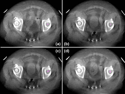
| Figure 2: Validation of deformable image registration between two CBCTs from the prostate fossa case. (a) Reference CBCT image with the tip of left femoral head marked (pink cross). (b) Sencondary CBCT image with the tip of left femoral head marked (pink cross). (c) The distance between the selected anatomical points before image registration. Points are overlaid on the secondary CBCT image. (d) Secondary image after deformable registration to the reference image, showing excellent match between two pink points. |