|
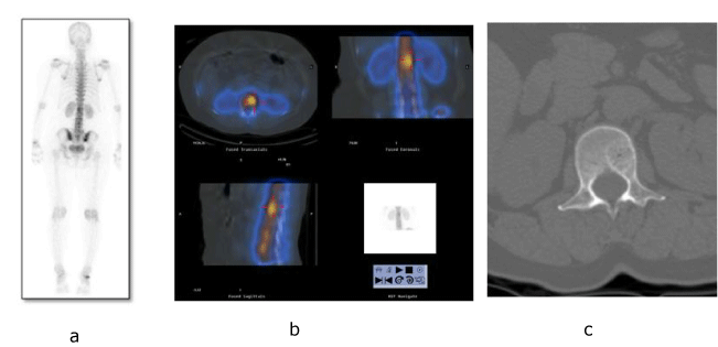
| Figure 1: a) Planar images of bone scan showing increased tracer uptake in L1 vertebra b) SPECT-CT fusion images revealed increased tracer uptake in L1 vertebral body corresponding to a osteolytic lesion in CT images C) CT section in bone window at L1 vertebra showing a well defined lesion with peripheral sclerotic rim and central ‘polka dot sign’. |