|
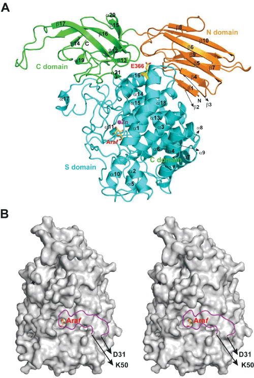
| Figure 1: The overall structure of HypBA1-L-Araf. (A) A cartoon model of the HypBA1-L-Araf complex structure. Each domain is presented in different color: N-, S- and C-domain are in orange, cyan and green colors, respectively. The Zn2+, ion is shown as a magenta sphere, the bound L-Araf molecule and Glu366 is shown as yellow stick model. (B) The obvious difference between the native, apo and ligand-bound HypBA1 is the capping loop (colored in magenta). The surface representation of the complex structure by omitting the capping loop clearly shows that the loop covers the active site cleft upon ligand binding. |