 |
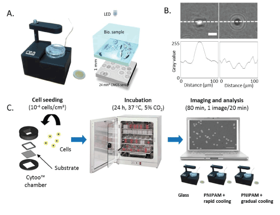
| Figure 2: Cell detachment monitoring and set-up. A. Photograph of lens free video microscopy (left) and schematic diagram illustrating its underlying principle (right). B. Difference in lensfree holographic patterns obtained from a cell while it is adhered to the substrate (left) and while it is floating (right). The scale bar is 20 μm. The images were acquired at 20-min intervals. When adhered to the substrate, the cell presents a holographic pattern with a bright center with a gray-value greater than 200 gray levels on a scale of 0-255. When the cell detaches from the substrate, there is a sharp change in the center of the holographic pattern, and the gray-value reduces to less than 100 gray levels. C. Protocol schematic of a cell detachment experiment and monitoring using lens free video microscopy. |