|
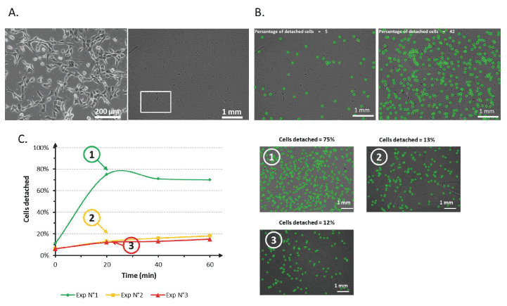
| Figure 4: Lens free imaging of cell detachment. A. Comparison of the field of view between 10X-magnified standard microscopy (left) and lens free video microscopy (right). The white square represents the size of the field of view of standard microscopy. B. Cell detachment quantification based on the data recorded via lens free video microscopy. Detached cells were identified using pattern recognition and are encircled in green. The percentage of detached cells was calculated at t=0 (left) and t=20 min (right). C. Cell detachment efficiency on a PNIPAM substrate with successive thermal detachment experiments. Cells were cultured in “red” media and were subjected to a “rapid” cooling. Images show the number of detached cells after 20 min of thermal-induced detachment for experiments n°1 (1), n°2 (2) and n°3 (3). Number of cells analyzed per experiment: 1851 +/-590. |