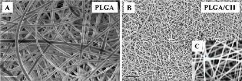
Figure 1:
Magnification of the structure of electrospun (A) PLGA and (B and C) PLGA/CH nanofibers visualized by scanning electron microscopy (SEM). Scale bars represent 10 µm in A, B and 1 µm in C.