|
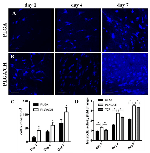
| Figure 4:Proliferation and metabolic activity of MSC seeded onto PLGA and PLGA/CH nanofibers. MSC nuclei were stained with DAPI (blue) on (A) PLGA and (B) PLGA/CH scaffolds at 1, 4, and 7 days post seeding. Scale bars represent 100 μm. (C) Proliferation measured as total cell count of MSC seeded onto PLGA and PLGA/CH nanofibers at 1, 4, and 7 days in culture. Cell proliferation increased up to 7 days post seeding on both PLGA (black) and PLGA/CH (white) scaffolds. The total cell count of MSC on PLGA/CH was significantly higher at all of the investigated time points compared to PLGA (p<0.05). (D) Metabolic activity measured as a reflection of mitochondrial function using an MTS assay in MSC seeded onto PLGA (black) and PLGA/CH (white) nanofibers and tissue-culture plastic (TCP, gray). Metabolic activity of MSC was significantly increased when growing on PLGA/CH compared to PLGA (p<0.05) and TCP (p<0.05). |