|
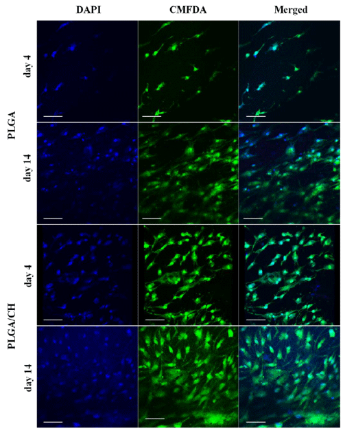
| Figure 5:Morphology of MSC seeded onto PLGA and PLGA/CH nanofibers after 4 and 14 days in culture visualized by confocal microscopy. MSC are labeled with a cell tracking dye (CMFDA) here seen in green and nuclei stained with DAPI (blue). At 14 days post seeding the MSC stretched out to over 100 µm and the total number of cells covered the surface at a higher density on both nanofibers. Scale bars represent 100 µm. |