|
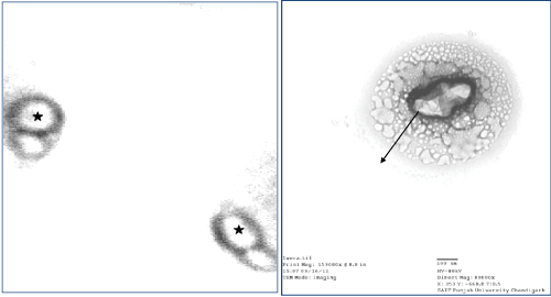
| Figure 1: TEM analysis of Alcaligenes sp. membrane vesicle after negative staining. TEM analysis of Alcaligenes sp. after 48 h of inoculation. Asterisk represents large PHB granules. Alcaligenes cell filled with granules. Arrows point to intracellular granules present inside membrane vesicle. Arrow represented the large PHB granule inside the vesicle (magnification 100 nm). |