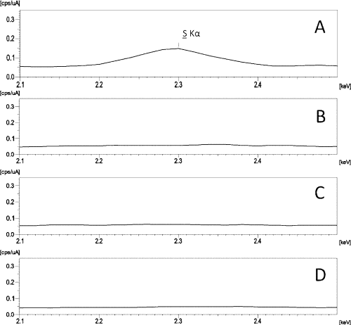
Figure 7:
Amplification of the spectrum of energy dispersive X-ray fluorescence showing the reduction in the concentration of sulphur observed in the sponge (A) for the purified silica Method I (B), Method II (C) and commercial silica (D).