|
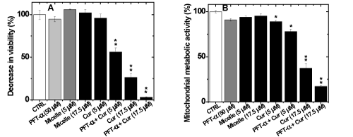
| Figure 7: 96-hour treatment with curcumin incorporated in micelles alone and with co-treatments significantly increased human glioblastoma cell death. Cells were then fixed and stained after treatment with the fluorescent Hoechst dye (10 µM, blue). Images were taken directly from the plate using Operetta imaging system (Perkin Elmer) and cell number was analyzed using the Harmony software. (A) U251N human glioblastoma cells co-treated with pifithrin-α (PFT-α 50 µM, 4 hours) and curcumin in micelles (Cur, 92 hours) ((PEG750)2–PCL4700 ) or curcumin in micelles alone (96 hours). Cell viability was analyzed by quantification of Hoechst stained nuclei. (B) U251N human glioblastoma cells co-treated with pifithrin-α (PFT-α 50 µM) and curcumin in micelles (Cur) ((PEG750)2–PCL4700 or curcumin in micelles alone were assayed for changes in mitochondrial metabolic activity using the MTT assay. Cells were treated with: 2 different concentration of curcumin in micelles (5 or 17.5 µM) for 96, 92 hours along with 0, 4 hours of pifithrin-α, respectively, for a total treatment time of 96 hours. (n=2). Statistically significant differences from untreated control were calculated using a t-test of OriginPro software and indicated by * (p < 0.05) and ** (p < 0.01). |