 |
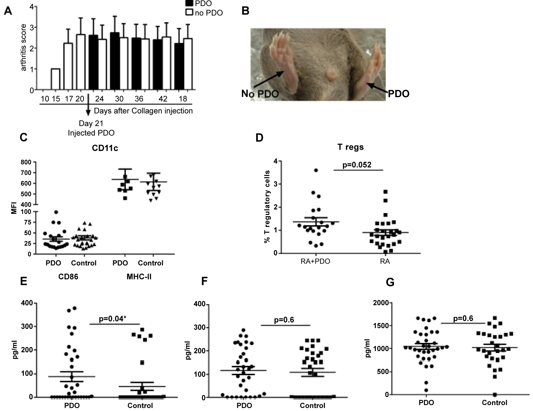
| Figure 4: Effect of PDO on inflammation in a murine model of rheumatoid arthritis. RA was induced in mice by injecting with collagen in CFA. 21 days later, 3 fibers of PDO were injected in the right thigh of mice. 4 weeks after the injection of PDO, the inguinal lymph nodeswere removed and the effect of PDO on DC activation, T reg generation and cytokine secretionwas measured using flow cytometry and ELISA. Left thigh lymph node from the same mice wasused as control. A. Bar graph depicts the arthritic score of mice in PDO- treated and untreatedlegs. B. Picture shows arthritic paw from PDO treated and non-treated mice at day 42 aftercollagen injection. C. Dot Plot depicts the MFI of expression of activation markers, CD86 andMHC-II on CD11c gated DCs. D. The percentage of CD4+, CD25+, FoxP3+ T regulatory cellswere measured by flow cytometry and are depicted in the dot plot. E, F & G. Collected lymphnodes were stimulated overnight with PMA and ionomycin and secretion of cytokines, IL-10,TNF-α and IFN-γ was determined by ELISA. Data is mean +/- S.E. of 28 mice. Each dotcorresponds to one mouse. |