|
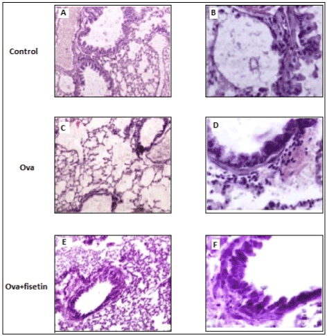
| Figure 20: Hematoxylin and Eosin staining of lung sections. Lung tissues were fixed in 4% paraformaldehyde overnight at 4°C, embedded in paraffin, cut into 5 μm sections, stained and observed under light microscope (Olympus). A, C, E: Lung sections observed under 10X magnification; B, D, F: sections observed under 40X magnification. |