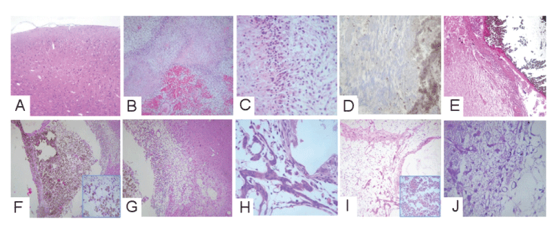
3a- tumor model used with Cu(NH4)2 Cl4 /F-TiO2. 3b-Tumor model used with Cu(Oac)2/F-TiO2 .3c-Tumor model used with Cu(acac)2/F-TiO2.
 |
| Figure 5: Histopathological findings. (a) Normal cortex, (b) now tumor shows
pleomorphic astrocytes, (c) nuclear pleomorphism, with mitotic activity center
necrosis around the vessels. In group (3a) are observed: (d) dense deposition
of the nanoparticles and (f) tumor around those particles. In group (3b) are
evidenced: (g) abundant macrophages immersed in nanoparticles and (h) the
peritumoral area with edema and gliosis. In group (3c) (i) the tumoral areas, that
the scarce neoplastic astrocytes are immersed in edema and vessels, in other
areas are presented polimorfonuclear leucocytes with necrosis and in (j) we
show inflammatory cells and gliosis. 3a- tumor model used with Cu(NH4)2 Cl4 /F-TiO2. 3b-Tumor model used with Cu(Oac)2/F-TiO2 .3c-Tumor model used with Cu(acac)2/F-TiO2. |