|
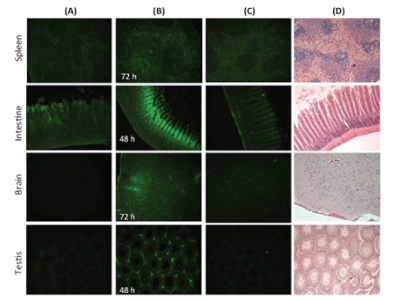
| Figure 3: Fluorescence microscopy images showing the distribution and localization of CdS-MDx QDs in tissues from rats. The images belong to brain and testis from male rats treated with a single dose of 200 μg/Kg of QDs. Tissue samples were analyzed under fluorescence microscopy at 20X. Column A shows tissues at 7 h, the first time of analysis; column B shows images of tissues observed at the time of maximal concentration (Tmax); column C shows images of tissues at 360 h; column D shows tissues stained with H&E at 360 h. The distribution and localization of CdS-MDx QDs was identified by a bright green imaging in the analyzed tissues. |