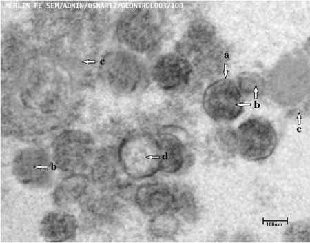
Size: 80-90 nm (a)
Shape: Typically cup and saucer (b)
Exosomal vesicles: Present (moderate) (c)
Electron translucent: Present (d)
Exosome collection speed pellet at 110, 000g
Sedimentation with sucrose gradient (0.25-2.5 M)
Scale bar 100 nm.
The 80- to 90-nm-sized microvesicles displaying the typical cup or saucer –shaped morphology of exosomes with moderate and translucent appearance in control group.