|
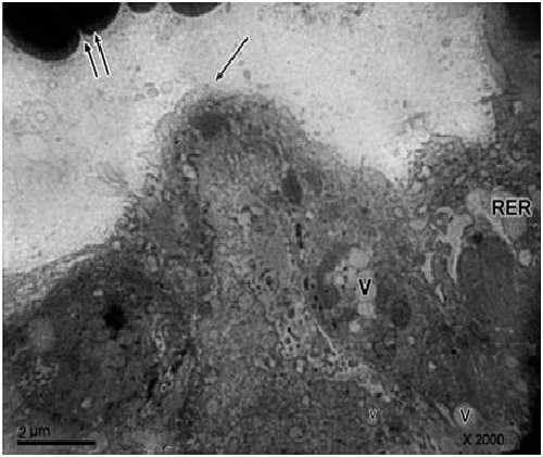
| Figure 9: Electron micrograph of a portion of a villous from preeclamptic placenta 14 days showing the maternal erthrocytes (↑↑) and irregularity of syncytial covering borders (↑).Notice the syncytiotrophoblast appeared with vacuolar cytoplasm (V)and dilated R.E.R.X2000. |