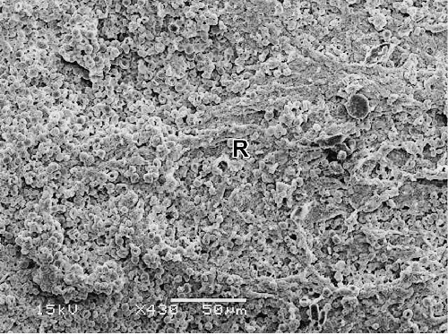
Figure 21:
A scanning electron micrograph of chorionic villi of pre-eclamptic placenta, showing the framework of preeclamptic villi become irregular, rough and thickened due to fibrin deposition. (×430).