|
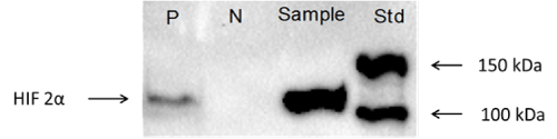
| Figure 2: Western blot image for positive control (P), negative control (N), sample (nuclear extract from kidney of 1-hour hypoxia treated rat) and molecular weight ladder (L) demonstrates the HIF-2α (118 kDa) can be detected using the specific HIF-2α antibody and there is no cross-reaction with other proteins, (N=4). *Values significantly different compared with the control group (* P < 0.05, ** P < 0.01, *** P < 0.001). |