|
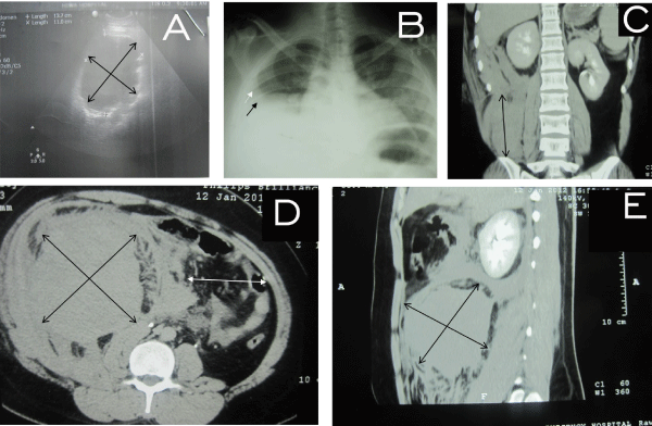
| Figure 1: A) Ultrasound of abdomen, in 22 years old man showing large hematoma (double arrows). B) Plane X-ray shows elevation of the RT dome of diaphragm (black arrow) with mild plural fusion (white arrow). C) CT scan show large hematoma in the right side of abdomen pushing the right kidney laterally. D and E) CT scan with intravenous contrast show large 150 × 120 × 100 mm right side retroperitoneal heterogeneous space occupying lesion (double arrows). The haemotoma was almost occupied the right side of the abdomen and pelvis associated with fat stranding in the retroperitoneal region. D) The mass was displacing bowel loops to the contra lateral side with contrast features of RPH (white double arrow). |