|
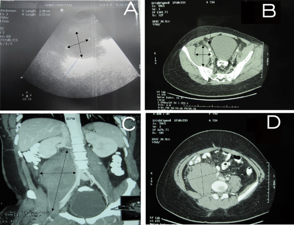
| Figure 2: A) Ultrasound showed large right sided pelvic collection about 200 × 80 × 70 mm (double arrows) extending to the right loin region. B, C, and D) CT scan of the abdomen and pelvis showed heterogeneous mass of 250 × 96 × 85 mm (double arrows in B and C, lines in D) not enhanced by contrast retroperitoneal in the right side of psoas muscle. |