|
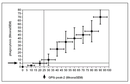
| Figure 3: GPIb clustering-like appearance (GPIb peak 2) versus aggravation of phagocytosis of C22 platelets after 0, 40 min, 48h and 72 h storage. The data presented are the Mean ± SEM of 20 different experiments in vitro . This figure illustrates the PCs based on quantitative GPIb-decay (-dependent) phagocyted by macrophages with a threshold of 25% (n=30). Hence, GPIb degradation and changes could be used as biomarker of the recipient’s immune response. |