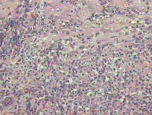
Figure 2:
Histological analysis of the lesion which shows the presence of leukocytoclastic vasculitis and an inflammatory infiltrate compatible with Sweet’s Syndrome diagnosis (Hematoxiline Eosine stain 40X).