 |
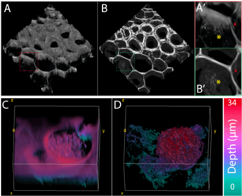
| Figure 3: STA for thick DIC imaging. 3D representation of Convalaria root slice directly (A, A’) or after STA (B, B’ and supplementary figure 3B). Direct reconstruction result in a strong loss of detail such has vacuoles envelops (yellow star) or cell wall (red cross) which are realistically preserved after STA. stack size: 1004 × 1002 × 163 voxels, voxel size 115 × 115 × 195 nm. Depth-color-coded 3D representation of membranes of plated MDA cells surrounding an apoptotic one, directly (C) or after STA (D). For such objects, no appropriate threshold can be found to discriminate between cell and background, thus direct 3D reconstruction is less informative than the optimal 2D DIC image. After STA, one can easily identify cell structure at different depth. In this example, membranes from different healthy plated cell (color-coded in blue to purple) are surrounding a thick round cell (color-coded from blue to red). This representation is of major interest for dynamic study of processes strongly affecting cell shape such as cell division or death where traditional 2D acquisition result in out-of-focus images of the studied events. Stack size: 422 × 481 × 62 voxels, voxel size 72 × 72 × 350 nm. |