|
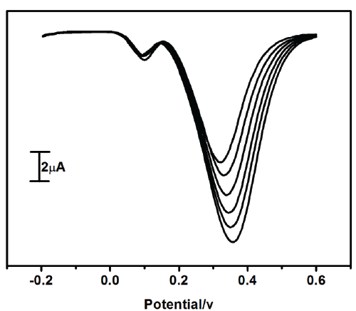
| Figure 8: Differential pulse voltammograms of uric acid different concentration (from a to e): 10 μM, 20 μM, 30 μM, 40 μM and 50 μM UA in Phosphate buffer solution at pH 7.5 in presence of 20 μM Dopamine at Pregabalin modified carbon paste electrode with a scan rate 100 mV/s. |