|
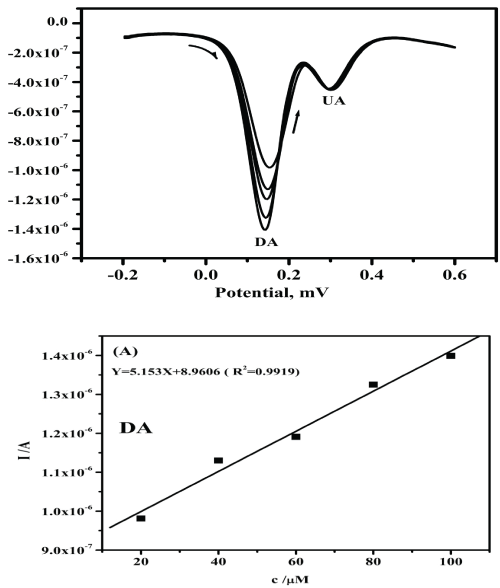
| Figure 10: Differential pulse voltammograms of DA (20 μM, 40 μM, 60 μM, 80 μM, 100 μM) in 0.2M phosphate buffer solution of pH 7.0 in the presence of 0.1 mM UA at GMMCPE with the scan rate of 100 mV/s. (a) The plot shows anodic peak current (Ipa) versus DA concentration. |