 |
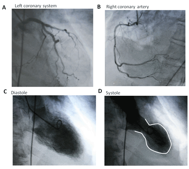
| Figure 2: Cardiac catheterization images did not demonstrate any significant disease in the left anterior descending and circumflex system (panel A) and right coronary artery (panel B). Left ventriculogram from the right anterior oblique projection at the end of diastole (panel C) and systole (panel D) demonstrated severe hypokinesis of the inferoapical and anteroapical wall (apical ballooning) with preserved basal segmental wall motion consistent with TC. |