|
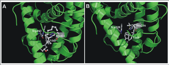
| Figure 5: Ribbon diagrams with the orientations of vitamin D3 and hydroxyl group interacting tryptophan and histidine residues (Trp443 and His421 in normal LXR-α protein (A) while Trp238 and His216 in patient LXR-α protein (B) in ball-and-stick mode at the ligand binding cavities are shown. The hydrogen bond between vitamin D3 and histidine residue of the ligand binding domain is shown by yellow color dotted line. In each figure the tryptophan and histidine residues are labeled by three-letter amino acid codes followed by the residue numbers. The vitamin D3 is labeled as vitD. |