 |
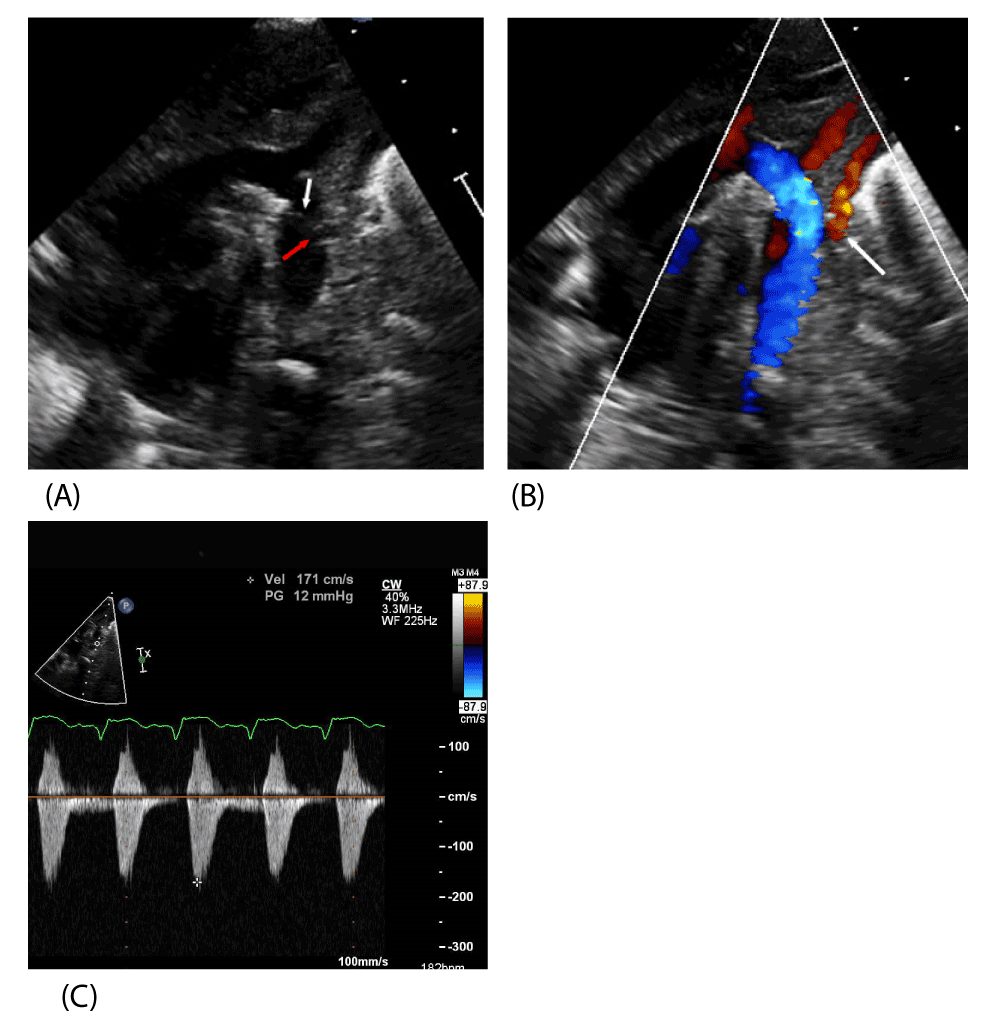
| Figure 2: A. 2D TTE performed after coarctation repair. The origin of the LSA is widely patent (red arrow). The arch repair is widely patent and the suture line is clearly visible (white arrow). B. 2D TTE with color Doppler interrogation demonstrating laminar flow in the left subclavian artery (white arrow). B, C. Color Doppler snows laminar flow in the descending aorta and spectral Doppler velocities are normal indicating no residual coarctation. Key: 2D TTE – two dimensional transthoracic echocardiogram, LSA – left subclavian artery. |