|
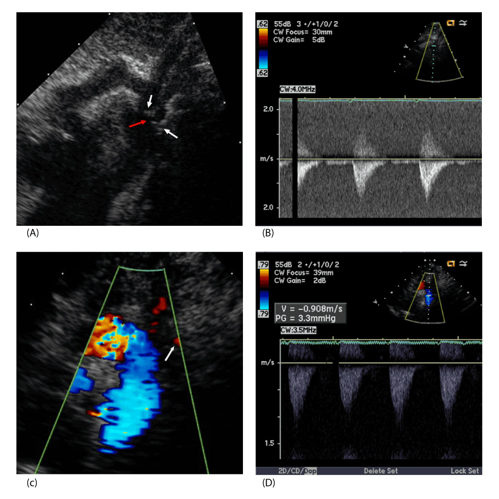
| Figure 3: A. 2 D TTE demonstrating the coarctation with involvement of the left subclavian artery. Note that as the posterior coarctation ledge (white arrows) extends into the aortic lumen that it includes and narrows the origin of the LSA (red arrow). B. Spectral Doppler interrogation demonstrates antegrade flow in diastole but no gradient with the PDA open. C. Post operative 2D TTE with color Doppler interrogation shows flow in the region of the LSA (white arrow). C, D. Color Doppler snows laminar flow in the descending aorta and spectral Doppler velocities are normal indicating no residual coarctation. Key: 2D TTE two dimensional transthoracic echocardiogram, LSA left subclavian artery. PDA patent ductus arteriosus. |