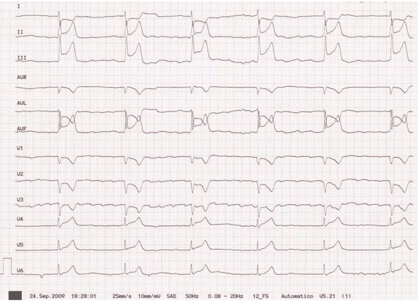
Figure 2:
ECG obtained at the onset of chest pain showing ST-segment elevation in inferior leads and reciprocal ST-segment depression in leads I and aVL.