|
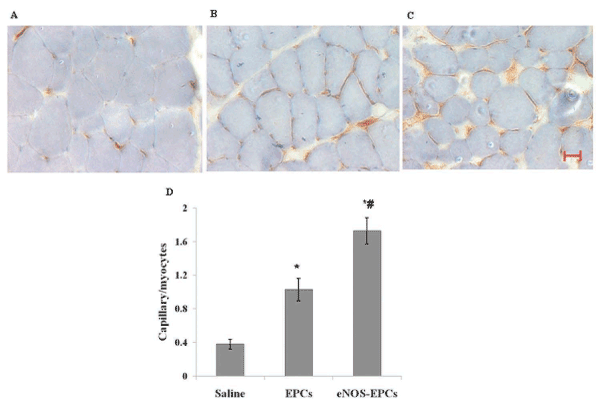
| Figure 3: Immunohistochemical staining of CD31 in ischemic hind limb muscle in (A) PBS treated (B) EPC treated (C) eNOS-EPC treated group. Brown spots are CD31-positive cells. Blue represents cells counter-stained with hemotoxylin. Scale bar represents 30 µm. (D) Histogram showing the mean capillary density as the ratio of CD31-positive cells to total no. of myocytes in five random fields from two muscle sections of each animal (n= 6 in each group). Results are expressed as mean + SD. *P< 0.05 versus PBS treated group and #P= 0.05 versus EPC treated group. |