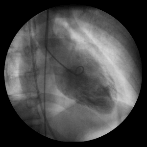
Figure 3:
Left Ventriculogram- motionless, depicting normal apical wall anatomy with the absence of apical ballooning.